

What do vision, audition, touch, and the other senses have in common?
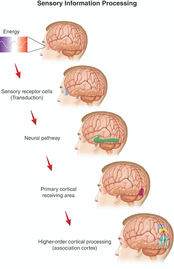
Vision begins when the light reflected off of objects (trees, tables, computers) hits sensory receptors in the retina of the eye. But at the very moment those visual sensory receptors (photoreceptors) respond to light, we have not yet had the inner experience of 'seeing' the object. The conscious perception requires that the photoreceptors activate neurons that carry the visual information toward the brain (see "neural pathway" in the figure). The subjective visual experience is believed to occur when the visual information, reaches the primary cortical receiving area less than a tenth of a second later. For vision, that cortical receiving area is the primary visual cortex. When activity in the primary visual cortex (called V1) is disrupted, individuals lose conscious visual awareness. From the primary cortical receiving area, sensory information is sent to high-order regions that give rise to things like perception of an object's color, its location in space, even its emotional significance to the viewer.
Similarly, auditory stimuli, carried by sound waves, reach sensory receptors (hair cells in the ear), and the auditory information travels along a neural pathway that transmits the signal to the primary auditory cortex, the auditory system's primary cortical receiving area where hearing occurs. Transmission of the auditory signal to the auditory cortex is even faster than that of the visual information to the visual cortex.
Sensory receptors: Turning energy 'out there in the world' into neuronal responses
When we speak of turning light waves, sound waves, and other kinds of detectable external energy into neuronal responses, we are speaking of transduction. Sensory processing begins with transduction. Transduction is what sensory receptor cells do. Let's consider the photoreceptors in the retina of the eye.
The first thing to notice is that the retina is at the back of the eye, and the photoreceptors are located at the back of the retina. Light entering the eye has to pass through several other cell types before it reaches the light-sensitive photoreceptors. You might wonder whether those other cells (ganglion cells, bipolar cells) block much of the light traveling to the photoreceptors. But they don't, largely because those other cells are highly transparent.
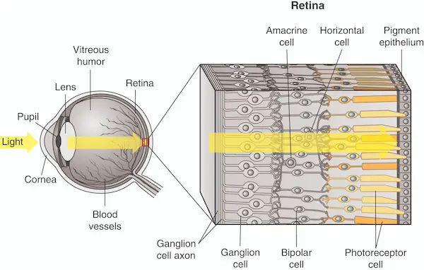
So, if you're looking at, say, a tree, the light carrying the image of the tree (that is to say, the light reflected from the tree) reaches the photoreceptors largely unimpeded. The photoreceptors detect that light and change their firing rates as a result. That is transduction, a change in neuronal activity in response to physical energy; in this case, light. If we call this change in photoreceptor firing rate the 'signal', then we can say that the photoreceptors send this signal to other neurons that transmit it to the brain through a particular 'pathway', which we'll consider next. But first, let's review.
Test yourself
These six steps describe what happens between light reaching your eye and a fully processed visual experience. They're shown here out of order. Assign each one a number (1 through 6), then click Check Order.
From the retina to the brain
We've seen that photoreceptors, the visual system's sensory neurons, respond to light upon the retina. But we'd like to know about the two main types of photoreceptors and how they differ. We'd also like to know how the photoreceptors send a visual signal to the brain.
Take a few minutes to read about the terms listed below. If you're using Principles of Behavioral neuroscience, you'll find them in the following sections of chapter 3.
Section 3.2.2: Photoreceptors: The light-sensitive neurons of the retina
- rods
- cones
Section 3.2.3: From light to retinal output
- bipolar cells
- retinal ganglion cells
- lateral geniculate nucleus of the thalamus
Go over these, and then come back to test and consolidate your understanding. By the way, besides the rods and cones, there is a third type of photoreceptor. Once you've consolidated your knowledge with the test-yourself questions below, I'll tell you about that third type that isn't mentioned in the textbook. Aren't you glad you found this tutorial?
Practice until you own it
1. Which are photoreceptors?
2. Which are highly sensitive to even dim light?
3. Which respond differently depending on the color of the image (wavelength of light).
4. Which provide high visual acuity (allowing objects to be perceived in detail)?
5. Which of these neurons is located in the retina?
6. Which of these neurons has an axon that leaves the retina and terminates in the brain?
7. Which of these neurons 'transduces' light (which carries out transduction)?
Test yourself
Let's ask again about the order of steps that occur beginning with light's arrival to the eyes. This time in more detail. The steps are shown here out of order. Assign each one a number (1 through 5), then click Check Order.
I told you I'd mention the 3rd type of photoreceptor that gets much less attention than the rods and cones. It gets less attention because it doesn't contribute directly to our visual experiences. These photoreceptors are called intrinsically photosensitive retinal ganglion cells. One of their functions is to adjust the size of the pupil in response to changes in light intensity. When bright light enters the eye, the pupil gets smaller, protecting the retina from light overexposure. Another role is to signal to the biological clock that morning has arrived (chapter 5: Sleep, in Principles ...). Notice that neither of these functions involve processing visual images that reach the retina and give rise to visual experience. That's the job of the rods and cones.
Filling in the missing pieces
Here are some additional things that you'll want to explore in order to give yourself a strong background in the neural basis of vision and other sensory systems. In Principles of ..., these are in chapter 3.
Priority 1- What do we mean by a neuron's "receptive field"? This is a key neuroscience concept.
- How do receptive fields in ganglion and LGN neurons differ from receptive fields in primary visual cortex (V1)?
- How was the receptive field of V1 neurons discovered? (Hint: it involves a glass slide.) If you understand this accidental discovery, you'll really get the idea of what's meant by a neuron's 'receptive field'.
- What are the dorsal and ventral streams of the visual system? What would your life be like with only the dorsal stream, or only the ventral stream? (Spoiler alert: it would be weird.)
- Blindsight
- Prosopagnosia All priority 1 topics are in sections 3.2.4-3.2.7 (1st edition); or 3.2.4-3.2.9 (2nd edition, 2027)
- What are meant by 'orientation columns' of the visual cortex? (I'm listing this as priority 2, compared to the priority 1 topics. But these 'orientation columns' were a major neuroscience discovery.)
- Synesthesia
- Sensory receptors for hearing (hair cells), touch (mechanoreceptors and nociceptors), smell (olfactory receptors), and taste (taste receptors)
- Cortical receiving areas for hearing, touch, smell, and taste. These topics are in 3.2.7 and 3.3 (1st edition) ; 3.2.9 and 3.3 (2nd edition)
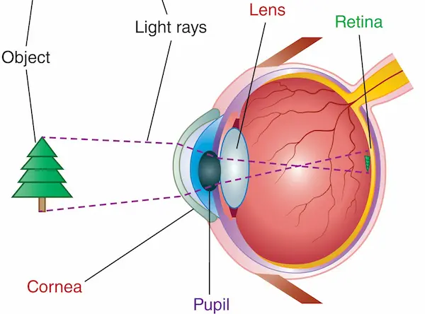
- The basic structure of the human eye (cornea, pupil, lens, retina)
- The fovea
- Right and left 'visual fields'; How is it that information from the left visual field is sent to the right visual cortex, and vice versa?
- Neural pathways between sensory receptors and cortical receiving areas for hearing, touch, smell, and taste These topics are in 3.2.1-3.2.3 and 3.3 ( 1st and 2nd editions)