

Where to start?
I think a good place to start is by touching your own hand. Really. Now realize that you didn't experience the sensation until the touch activated receptors in the skin, those receptors sent information to the spinal cord, and the spinal cord sent the information to the brain.
The touch receptors are called somatosensory receptors, and they come in different types. Some are sensitive to gentle touch, others to strong touch, still others to pain. The book discusses the different types of somatosensory receptors, but that's not key here.
What's interesting to think about here is that you don't feel the touch sensation at the moment that something makes contact with your skin. Instead, the 'feeling' arises from brain activity. So the brain needs to find out about the touch to your skin.
How does the touch information which begins at the somatosensory receptors in your skin get all the way to the brain? Neurons carry the information. And so understanding what neurons are, and how they carry 'information', becomes key to understanding any inner experience, including the experience of touch. We'll think about neurons a little later.
But before we get to neurons, let's briefly go through some key regions of the brain. We'll begin with the base of the brain (the brainstem), examine the brain areas below the cortex (subcortical regions), and finally reach the outer layers of the brain, the cerebral cortex, where sensations of touch, hearing, and vision occur.
The Brainstem: Functions critical for life
The brainstem includes the medulla, pons, and midbrain - in that order as you climb above the spinal cord and ascend into the brain.
- The medulla and pons are critical to basic survival functions, including regulation of breathing, heart rate, and blood pressure. Of all brain injuries, damage to the medulla and pons is the most life-threatening.
- The midbrain is important in several aspects of behavior, but of particular interest is the fact that most dopamine neurons originate (have their cell bodies) in the midbrain and send signals to motivation-related brain regions where they play a role in reward-seeking and addiction. The midbrain also contains two other key regions, the inferior and superior colliculi, described below.
To get a sense of the position of these brainstem structures, take a look below. A good trick for distinguishing the front from the back of the brain is to look for the cabbage-shaped cerebellum. That's always at the back of the brain.
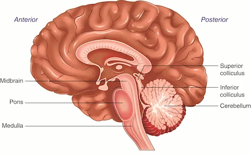
Notice that the three brainstem areas are labeled to the left in the illustration.
To the right, you'll see labels for the inferior colliculus and superior colliculus. Those are two regions of the midbrain that respond to sounds and sights, respectively. These midbrain areas only respond to events that are so obviously important (say a loud sound or the sudden appearance of a visual object near you) that they demand immediate attention - even before the cerebral cortex has had a chance to find out details about exactly what the auditory or visual event was.
On the bottom right, you'll also notice the large cabbage-shaped cerebellum attached to the back of the pons.
- The cerebellum plays a key role in movement coordination and the ability to learn skilled movements that require fine precision. Alcohol can disrupt one's balance and skilled movements in large part because it impairs cerebellar function.
Practice until you own it
1. Which brainstem structure is directly on top of the spinal cord?
2. What brain area is just above the medulla?
3. What brain area lies just behind the pons?
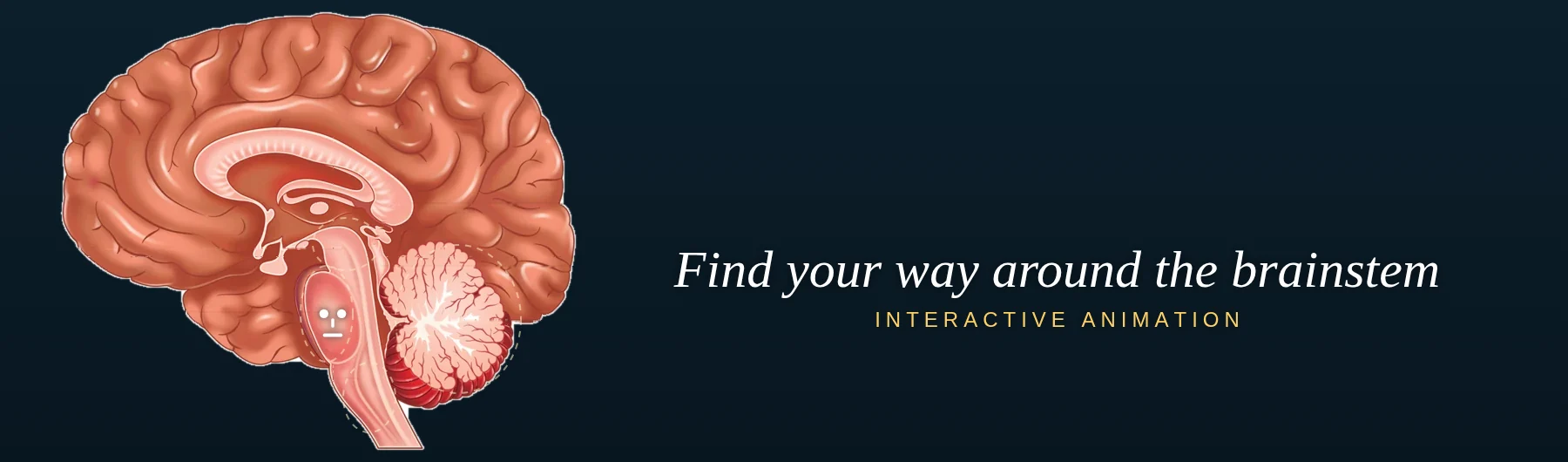
Try to answer questions 1 through 3 from memory, without looking at the brain image above. If you'd like a clean slate first, refresh this page; this clears any highlighting from your last attempt, so you won't see which answers you already picked.
Subcortical Brain Regions: Motivation, emotion, memory and other functions
If you think of the brainstem as the basement of the brain and the cerebral cortex as the penthouse, there are a number of important brain structures between the two. For now, we'll consider just five of the most important ones: the hypothalamus, thalamus, hippocampus, amygdala, and basal ganglia.
- The hypothalamus is critical for hunger, thirst, sex drive, and other innate drive states.
- The thalamus receives information from the senses (vision, audition, etc) and passes it on to the cerebral cortex which, in turn, gives rise to the experience of seeing, hearing, etc. It is sometimes referred to as a 'relay' station. For example, the neurons in the retina of the eye send visual information to an area of the thalamus that is dedicated to vision, and neurons originating in this thalamic nucleus relay the information to the visual cortex. When you wish to attend to a particular object in the environment, the cortex commands the thalamus to pass on more information about that object to the cortex. For instance, the cortex may decide that it wants to receive more information about the red object behind a bush, and commands the thalamus to send it more visual information about that object.
- The hippocampus plays a key role in memory. Individuals (and animals) who have suffered damage to the hippocampus are greatly impaired in storing and retrieving memories of events they've experienced.
- The amygdala is critical for learning about which environmental situations are dangerous. For instance, if you suffer a physical attack in a particular location, you will learn to be afraid when you pass by that location. Individuals (and animals) with amygdala damage fail to learn associations between environmental stimuli and aversive outcomes. A woman who suffered complete damage to the amygdala on both sides of her brain failed to acquire associations to dangerous stimuli, and showed virtually no signs of fear even in very dangerous situations.
- The basal ganglia are a group of brain structures that play an important role in allowing the behaviors that we frequently perform to become automatized habits. Since so much of our daily behavior involves acquired habits (using silverware, dialing the phone, getting dressed, etc., with little conscious attention required), a loss of basal ganglia function produces great difficulties in behavior. Usually, those with basal ganglia damage can still move, but movement becomes slow and extremely effortful.
Now take a look in Google to find images that include these brain regions. It's possible that you'll have to look at multiple images, for some brain illustrations will only show a subset of them. The idea is not to become a neuroanatomist today, but to get a feel for where they lie with respect to one another.
Because these five areas are below the cortex, they are called "subcortical areas." When we talk of subcortical areas, we don't usually include the brainstem, even though it is far below the cortex. Similarly, when someone says they're working on one of the lower floors of an office building, they probably aren't referring to the basement, even though it's the lowest floor. When you feel comfortable with the descriptions of these subcortical areas, try the questions below. They're straightforward.
4. Damage to this area can abolish the motivation to eat and drink.
5. As you recall what you ate for dinner last night, you are activating your _____.
6. This collection of brain structures is needed in order for actions that you repeat many times to become automatized habits.
Review of material so far (brainstem and subcortical regions)
Bob received a gunshot wound to the , an area of the brain housing several brain regions, including the medulla, pons, and midbrain. The injury didn't affect all of these regions. Fortunately, there was no damage to the that sits just above the spinal cord. However, he did suffer damage to the where his dopamine cell bodies (the origin of the dopamine neurons) are located. It's likely he'll survive, but it's possible he'll have particular problems with . If the damage were to his hippocampus, it's likely he'd have had problems with .
brainstem, medulla, midbrain, motivation, memory
Cerebral Cortex: Perception, Action, and Cognition
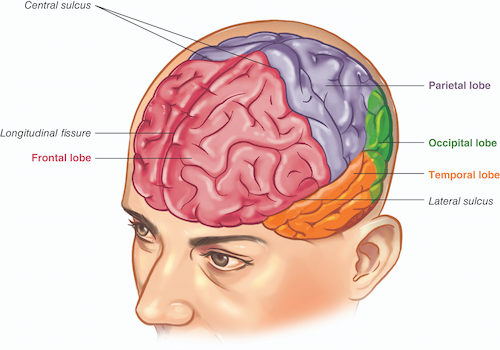
The cerebral cortex is comprised of lobes that are critical for vision (occipital lobe), audition (temporal lobe), touch or 'somatosensation' (parietal lobe), actions (frontal lobe) and cognition (several of the lobes, but especially regions of the frontal lobe). In this drawing, you'll notice that in addition to the four lobes, there are pointers to grooves 1) between the left and right hemispheres of the brain (longitudinal fissure), 2) behind the frontal lobe (central sulcus), and 3) above the temporal lobe (lateral sulcus). As you might have guessed from the drawing, the word we use for a groove within the cortical surface is sulcus (sulci, pl.) or fissure. The terms sulcus and fissure are sometimes used interchangeably.
I wouldn't worry about remembering the names of these sulci/fissures at this point. But you should remember that the term sulci (or fissure) refers to grooves in the cortex. You'll notice that the illustration shows many sulci in the cortical surface, with only a few labeled. Also, notice that between sulci, the cortex bulges outward. Each of the bulges between sulci is called a gyrus (gyri, pl.). Take a moment to Google some images for 'sulcus' and 'gyrus' and point to some of the sulci and gyri so you become accustomed to their appearance.
Notice that a few paragraphs above, I said that the central sulcus is behind the frontal lobe. Neuroscientists rarely use the terms 'in front of' or 'behind', but instead say anterior (toward the nose) or posterior (toward the back of the head). So, as you can see in the illustration, the frontal lobe is anterior to the parietal lobe. The central sulcus and the parietal lobe are posterior to the frontal lobe. Sometimes, instead of anterior vs posterior, you'll see rostral vs caudal. Finally, instead of saying above vs below, we say dorsal vs ventral. So, if you look at the illustration of the cortex, you'll see that the lateral sulcus is dorsal to the temporal lobe, and the temporal lobe is ventral to the lateral sulcus. At the end of this tutorial, you'll see suggested terms and concepts to look up on your own -- they'll include terms that point to particular directions within the brain.
Review of material so far (cortex, subcortical, and brainstem structures)
You are looking at a tree and the visual experience is due to activity in the lobe. The scene reminds you of a childhood memory which comes to mind thanks to activity of your . You walk to the tree and raise your arm toward a branch. The movement of your arm requires that you activate neurons in your lobe which generates actions. Suddenly you hear a sound. Your immediate detection of the sound, before you've had a chance to process the details of what the sound is, involves activation of neurons in an area of the midbrain called the superior . A brief moment later you recognize the sound to be the song of a bird. The auditory perception required activation of neurons in your lobe.
hippocampus, colliculus, occipital, temporal, frontal
Taking it to the next level
For students: Here are some additional things that you'll want to explore in order to give yourself a strong overview of the brain and the rest of the nervous system. The terms and concepts below are described in Chapter 1. Understanding them will give you a good background for the more specific topics we'll explore in later chapters:
Priority 1- The basic parts of a neuron: cell body, dendrites, axon, terminal. You really want to 'own' these terms. They'll come up again and again in later chapters, and you don't want them to read like jargon.
- Neurotransmitters, receptors, and the synaptic cleft. For now, just get a feel for what they are. You'll learn more about them in the next chapter.
- Central nervous system vs peripheral nervous system This is the key division within the nervous system
- Sympathetic nervous system vs parasympathetic nervous system This is the key division within the autonomic nervous system. You are probably familiar with what it feels like to have an active sympathetic nervous system, especially if you're someone who feels stress and anxiety often.
- Terms that allow you to describe the location of brain regions. If you know how far anterior-posterior (toward the nose or back of the head), dorsal-ventral (toward the surface or the base of the brain), lateral-medial (toward the ears or the midline) a brain site is, you can pinpoint exactly where it is located in the brain
- Corpus callosum, which connects the left and right sides of the brain
- From this tutorial, you already have an introduction to the brainstem (medulla, pons, midbrain), cerebellum, thalamus, hypothalamus, basal ganglia, amygdala, hippocampus, and the outermost part of the brain, the cerebral cortex, specifically, its areas within the frontal lobe, parietal lobe, temporal lobe and occipital lobe. In Chapter 1, you can expand your knowledge of these brain structures.
- Glia. While listed here as Priority 2, many researchers dedicate their professional lives to understanding glial cell function. The chapter describes a study where a particular type of glial cell (the astrocyte) from a human brain was implanted into the brains of mice. It reads almost like science fiction.
- Grey matter and white matter. The brain can gain and lose grey and white matter over your lifetime. What exactly is being gained or lost?
- Afferent vs efferent nerves. The brain receives information from the senses, allowing you to see, hear, etc., and even receives messages from the body itself -- allowing the brain to know when your heart is racing, or when your muscles have tightened up. The brain also sends information out to the periphery of the body to activate muscles. It's good to be clear on the distinction between information entering versus leaving the brain. Figure out which is afferent information and which is efferent information -- they're useful terms to know.
- cranial nerves
- ventricles
- pituitary gland
- meninges
- brain imaging approaches
- pineal gland
Remember, the Student Resources page provides additional study material, suggested readings and videos, answers to the textbook's Test Yourself questions, and neuroscience-related blog posts.