


Neurons communicate
Even if you're not familiar with how neurons communicate, I'll bet you've heard people speak of one neuron sending a 'message' or a 'signal' to another neuron. Maybe you've even read things like
When Sarah saw a huge bear approaching her camping tent, neurons in her amygdala were activated. As a result, she became so scared that her heart rate went through the roof.
The phrase suggests that neurons that responded to the sight of the bear activated her amygdala. Neurons in her amygdala generated fear and communicated with her heart muscles, causing an accelerated heart rate. That's all true. In fact, the amygdala neurons also would likely have sent signals to other neurons to activate sensory regions of her cortex, making her highly vigilant to sounds and other sensory stimuli that might signal danger. Neurons in the amygdala communicate with many brain regions that together give rise to bodily responses and an inner experience of fear.
Maybe Sarah thought to herself "Okay, try to relax. The bear is still far away, and the chances are that it will just pass by." If her reassuring thoughts were enough to allay her fears it is likely because some 'thought-related' neurons in certain parts of her prefrontal cortex were able to inhibit the amygdala neurons that were generating the fear. It doesn't necessarily mean that the thought-related neurons had direct connections with the amygdala neurons. The connection may have been indirect. For instance, let's call the thought-related neurons 'A'. Some of those A neurons may have had connections with other neurons (call them 'B'), which in turn had connections with the amygdala neurons ('C'). It could be that the 'A' neurons activated the 'B' neurons, and the 'B' neurons suppressed activity of the 'C' neurons. In other words, the thought-related A neurons indirectly suppressed activity of the C neurons in the amygdala that were activated by the sight of the bear.
Two things are key in the scenarios above. First, in order for the activity of one neuron to affect another, the two neurons have to be physically (anatomically) connected, either directly or indirectly. Second, some neurons are able to excite their recipient neurons, while other neurons produce inhibition. In some cases, a neuron has effects that can't be described simply as excitatory or inhibitory. But in most cases, a neuron is either excitatory, causing its recipient neuron to fire more, or inhibitory, causing its recipient neuron to fire less.
There's actually another key issue that arises from the examples above. Is it the activity of neurons that ultimately generates emotions, like Sarah's fear from seeing the bear, and thoughts, such as those that may have reduced Sarah's fear? Does neural activity give rise to emotions and thoughts, or do emotions and thoughts produce changes in neural activity? Perhaps it's not a question of one or the other, but both are occurring at the same time. How should we think about this relationship between the neuronal and the mental? Philosophers and neuroscientists have given a lot of thought to this. The intimate connection between neuronal and mental activities comes up again and again in the chapters of Principles..., and in these interactive tutorials.
What do we mean by 'communicate'?
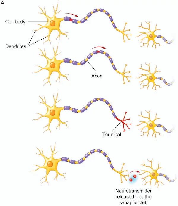
This is the question I was secretly hoping you'd ask yourself. There is something unsatisfying about just saying that one neuron 'communicates' with another, isn't there? It's a shorthand way of speaking. But the lack of detail actually makes the idea of neural communication more difficult to understand, not less.
Here we're looking at one neuron (on the left side of the drawing) communicating with another neuron (right). The branch-like dendrites receive information or 'input', in most cases from other neurons. Later, we'll ask what's meant by 'input'. If the neuron receives enough excitatory input, a signal will move along the long, thin axon, and toward the terminal where it releases neurotransmitter molecules into a gap between the two neurons called the 'synaptic cleft'. The cell body and dendrites of the receiving neuron contain receptors that detect the presence of the neurotransmitter.
Practice until you own it
1. A neuron receives information (often input from other neurons) at its branch-like:
2. The round portion of the neuron that collects the information from all the dendrites is the:
3. The signal moves along the long, thin _____ , which ends at the terminal.
4. When the signal reaches the terminal, a _____ is released into the synaptic cleft.
Neural communication in action: Dropping a hot cup
The signal moves from 1) the red neuron, to 2) the small spinal cord neuron, to 3) the green neuron
In the animation, the neuron activated by the heat of the cup is in red. You may recall that the dendrites of most neurons receive input from
other neurons. But the dendrites of sensory neurons in the skin respond to heat, pressure against the skin, tickle, and other tactile stimuli. The skin
contains different types of neurons to detect these different types of tactile ('somatosensory') stimuli. In the same way, neurons in the
eye respond to light; neurons in the ear respond to sound, and so on for the other senses. Chapter 3 examines these in some detail.
Each of these types of sensory neurons releases a neurotransmitter that crosses a synaptic cleft to activate another neuron.
While the animation does not show
neurotransmitters being released, crossing a synaptic cleft, and binding to receptors on the dendrites of the receiving neuron,
try to picture this occurring as you watch the animation.
For instance, you may imagine the heat-sensing neuron in the hand releasing neurotransmitter to activate the spinal cord neuron.
Release of neurotransmitter from the spinal cord neuron activates the green motor neuron ('motor' because it leads to a body movement). The green
neuron sends a signal down its axon and releases neurotransmitter that activates muscles causing the hand to open.
That's when the cup drops.
Brain function depends upon the negativity inside of neurons
So far, we've said that a neuron receives input that can excite the dendrites, and these excitatory signals are 'collected' in the cell body. Let's look at this more carefully.
![[Enter Figure]](images/Inside_outside of neuronA.webp) The inside of the neuron is negatively charged
compared to the outside. In the illustration, this is represented by the minus signs inside the neuron. The inside of the neuron is filled with fluid,
'intracellular' fluid. So is the outside of the neuron, the extracellular fluid.
So, we can say that the intracellular fluid is negative compared to the extracellular fluid. (We speak of intra- and extra-cellular fluid because a neuron
is a kind of cell.)
Notice in the figure that the neuron's 'membrane' is like a wall that keeps the intracellular fluid separate from the extracellular fluid.
The inside of the neuron is negatively charged
compared to the outside. In the illustration, this is represented by the minus signs inside the neuron. The inside of the neuron is filled with fluid,
'intracellular' fluid. So is the outside of the neuron, the extracellular fluid.
So, we can say that the intracellular fluid is negative compared to the extracellular fluid. (We speak of intra- and extra-cellular fluid because a neuron
is a kind of cell.)
Notice in the figure that the neuron's 'membrane' is like a wall that keeps the intracellular fluid separate from the extracellular fluid.
![[Enter Figure]](images/Inside_outside of neuronB.webp)
In the bottom illustration, you can see what's really present inside and outside the neuron. Notice the "Prot-" inside the neuron, which stands
for negatively charged proteins. These represent very large molecules that have a negative charge. From the illustration, it may look like they are
only present in the cell body, but they fill the entire neuron, including the long axon.
These negatively charged proteins are so large that they can't pass through the membrane, so they can't leave. They're trapped inside the neuron.
Notice that the inside of the neuron also contains "K+". K stands for potassium. '+' represents a positive charge.
And any atom with a positive or negative charge is called an ion. K+ (the potassium ions) are shown inside the neuron because they are
more concentrated there. You might wonder whether their positive charge should cancel out the negative charge of the proteins (Prot-).
To a large extent, they do. However, some K+ is constantly leaking out of the neuron through the membrane. Each K+ ion that leaks out takes a bit of positive charge with it.
Since the negatively charged proteins are trapped inside and can't follow, this ongoing loss of K+ is what leaves a negative charge on the
inner face of the membrane (the part of the membrane in contact with the intracellular fluid), and makes the
inside of the neuron negative compared to the outside.
If the inside of the neuron were not negative compared to the outside, we'd lose our ability to see, hear, think, and
experience emotions. That may sound like an exaggerated claim. But as you'll see it's 100% true. Understanding that the inside of a neuron is negative compared to the outside
is fundamental to understanding the 'signal' that moves across the neuron and allows it to communicate with other neurons. The resulting neural communication is
what gives rise to our inner experiences.
We haven't covered the ions in the extracellular fluid yet, but first let's consolidate. You really want to know this material so well that
if someone wakes you at 3am and asks you what ions are located in the intracellular fluid, you'd automatically
mumble "positively charged potassium ions, abbreviated K+, and negatively charged proteins. Now let me get back to sleep."
Review of material so far
Parts of the neuron
A neuron has long thin branches called 1), which receive input from other neurons. Connected to these branches is the round 2), which, in turn, gives rise to a long, thin 3), which finally ends at the 4), and releases 5) molecules into a gap between neurons called the 6). When the molecules cross that gap, they bind to receptors on the dendrites (and sometimes on the cell body) of the next neuron. Notice that neuronal communication is one-way traffic: a signal moves from the dendrites to cell body (also called the 'soma') to axon to terminal to synaptic cleft, almost always in that order.
1) dendrites 2) cell body 3) axon 4) terminal 5) neurotransmitter 6) synaptic cleft
The intracellular fluid
The entire neuron (its dendrites, cell body, axon, terminal) is completely surrounded by a 1) which separates the inside from the outside of the neuron. The fluid inside the neuron is called 2) fluid, while the exterior side of the membrane is filled with the 3) fluid, which surrounds the neurons. The intracellular fluid contains large 4) that are unable to leave the neuron. In the drawings above, they are abbreviated Prot-. These large protein molecules contribute a strong negative charge to the inside of the neuronal membrane. But the intracellular fluid also contains positively charged 5) (spell it out), abbreviated 6). Overall the inside of the neuron is 7) compared to the outside.
1) membrane 2) intracellular 3) extracellular 4) negatively charged proteins 5) potassium ions 6) K+ 7) negative
Na+ in the extracellular fluid can flow through channels to enter the neuron
![[Enter Figure]](images/ion channels 500w.webp)
In the dropping-the-cup animation, the signal shown moving
along the neurons is essentially Na+ channels opening in the axon one after another, and Na+ flooding in at each point.
It's important to have in mind that, for the most part, Na+ can only enter the neuron when Na+ channels are open. For the most part, because
just as a few people will be able to sneak into a concert through an unofficial side entrance even when the main entrance is closed, some Na+ ions will be able to slip through the membrane to enter the neuron even
when the Na+ channels are closed. But not many.
Na+ entry into the neuron is key to neuronal communication. Without it, you'll experience no pain, no pleasure, no anxieties, no hopes, no memories, no thoughts.
How does lidocaine or novocaine prevent you from feeling pain when the dentist drills into your tooth? They prevent Na+ from entering into neurons. Na+ isn't the only
ion involved in neural communication, but, as you'll see, it's the 'star of the show'.
This raises the question: what makes the Na+ channels open?
The short answer is that for some neurons, say neurons with dendrites in your skin, physical energy like touch or heat against the skin opens the Na+ channels. If enough Na+ enters, the neuron becomes activated, sends a signal along its axon, and releases a neurotransmitter to a receiving neuron. The neurotransmitter binds to receptors on that neuron, causing its Na+ channels to open. Similarly, sound waves vibrate tiny bones inside your ear, which ultimately causes Na+ channels in auditory sensory neurons to open. Again, if enough Na+ enters, the auditory neuron is activated and neurotransmitter is released. We examine the details of how tactile, auditory and other sensory neurons are activated in chapter 3 of Principles ...., but for here, the key idea is just that for sensory neurons in your skin, ears, eyes, and so on, physical energy is what causes Na+ channels to open.
Now, spoiler alert: We'll see later that for most neurons, Na+ channels in its dendrites and cell body open in response to a neurotransmitter released by another neuron. But regardless of whether we're considering a neuron that receives physical energy from the outside world (sensory neurons) or neurotransmitter from another neuron (most neurons), what I'd like you to get very clear on to begin with are 1) the ions concentrated inside vs outside the neuron, 2) the negativity inside compared to the outside, and 3) the fact that Na+, with its positive charge, will rush from the outside to the inside when a Na+ channel opens. The following immersive animation will help you to picture this.
Exciting and inhibiting neurons
In the animation, you saw Na+ entering whenever a Na+ channel opened. But why does Na+ enter when a channel opens? You may have noticed that the 'membrane potential' changes as Na+ enters the neuron. But what exactly is the membrane potential, and why does it change when Na+ enters?
If you have Principles of Behavioral Neuroscience , look in sections 2.2.1 and 2.2.2, about 2.5 pages of text and a few illustrations. It will answer these questions. If you don't have the book, another textbook can work. One way or another, try to get comfortable with the following terms:
From section 2.2.1
- diffusion
- electrostatic (or electrical) pressure
- the sodium-potassium pump
From section 2.2.2
- the membrane potential
- the resting potential
- What does it mean to say that a neuron is polarized?
- What does it mean to say that a neuron has become depolarized?
Go over these, and then come back to test and consolidate your understanding.
1. A neuron is at -70 mV at rest. Which membrane potential below indicates the smallest depolarization?
2. Which membrane potential below indicates the largest depolarization?
3. When Na+ channels open, Na+ rushes from the outside to the inside of the neuron. This is due in part because Na+ is positively charged and therefore attracted to the negativity inside the neuron. This attraction is called:
Na+ moves into the neuron, in part, because of its electrostatic attraction to the neuron's negative interior, but also because Na+ is more concentrated outside the neuron. The movement of a substance from an area of higher to lower 4) is called 5). But if Na+ enters the neuron every time its channels open, won't the inside of the neuron soon be filled with Na+? How can we be sure that Na+ will continue to enter the neuron if the inside has become full of Na+? In order to ensure that Na+ always starts out with a higher concentration on the outside of the neuron, it is constantly pulled out of the neuron by the 6).
When we say that the inside of the neuron is usually negative compared to the outside, we are saying that the neuron usually has a negative 7). A neuron that's more negative inside than outside is 8).
At rest, neurons are polarized, with a resting membrane potential of about -70mV -- that's typical for most neurons. If a neuron's membrane potential is -70mV and some Na+ flows in, the membrane potential might now be say -68mV or -66mV. In this case, we say that the neuron has become slightly less polarized, or 9). If you take another look at the interactive animation above, you'll see that as Na+ flows through a channel to enter the cell body or dendrites, the neuron becomes depolarized. Not enough to bring the membrane potential from -70mV to 0, but in that direction, say from -70 to -65 mV.
4) concentration 5) diffusion 6) sodium-potassium pump 7) membrane potential 8) polarized 9) depolarized
You've seen that Na+ entry depolarizes the neuron. In the animation below you can open Na+ channels anywhere on a neuron, and watch how that depolarization influence the membrane potential at the place where the decision to fire is actually made.
Interactive animation debriefing: If you've played with the animation, you may have noticed a few things.
- To reach the firing threshold at the axon hillock, it helps to open not just a single Na+ channel, but Na+ channels at several different locations on the cell body and dendrites. This adding together of depolarization arriving from different locations is called spatial summation.
- It also helps to open a Na+ channel many times in rapid succession. This adding together over time is called temporal summation.
- While the animation shows you how spatial and temporal summation can combine to strongly depolarize the neuron at the axon hillock, there's something that's not shown here. Each time the neuron fires, Na+ channels along the axon open one after another. And the membrane potential undergoes a brief change at each point along the axon as those open, including at the axon hillock itself, where the animation's meter simply holds steady above threshold. This is reviewed below and described in detail in textbook sections 2.2.4 and 2.2.5.
Putting the pieces together
Imagine a neuron with dendrites in the skin of your hand that is sensitive to heat. Right now there is no Na+ entering (or perhaps only a trickle that slip through the membrane and are quickly expelled). If you measure the membrane potential, it will be about about -70mV. The neuron is at 10). Now you pick up a hot cup, which causes the opening of 11) within the dendrites. Na+ rushes in. The polarized neuron is becoming slightly 12) The 13) in the dendrites has changed from about -70 mV to, say -68mV. It'sa very small depolarization. If the membrane potential remains at this level, the neuron won't fire That is, it won't send a signal along its axon to a activate a receiving neuron in the spinal cord. It's also unlikely that you'll drop the cup, because if the spinal cord neuron doesn't fire, it won't be able to send a signal to the muscle in your hand that would cause you to drop the cup. But imagine that the cup was much hotter. Now, when you touch it, more Na+ channels open in the sensory neurons of the hand, more Na+ enters, and the membrane potential now becomes, say, -50, a much larger depolarization. Now you do drop the cup. What happened?
10) rest 11) Na+ channels 12) depolarized 13) membrane potential
What does it mean when we say a neuron 'fires'?
As we've seen, a neuron receives input at its dendrites and cell body, collects these inputs within the cell body, and sends output to another neuron at its axon terminal. As we've seen, the input can come from neurotransmitter molecules released by other neurons or, in some cases, from energy in the external world, such as the heat that provides input to heat-sensitive neurons in the skin. Let's work with this latter example to begin with, just because it's so vivid. This will give you the key concepts needed to understand neuronal activation, and will make it easy for you to understand how neurotransmitters can activate neurons as well.
The heat-sensitive neuron in the hand responds to the heat of a cup by opening sodium channels in its dendrites that allow sodium to flow in. This depolarizes the neuron, at least a bit. If there's enough depolarization, a signal will move along its axon, causing release of a neurotransmitter at its terminal. When a signal moves along an axon and neurotransmitter is released, we say that the neuron has fired. A key question then is, how much depolarization is needed in order for the neuron to fire? And what exactly is the 'signal' that's moving along the axon of a firing neuron?
Take a look at Section 2.2.3, and familiarize yourself with 1) firing threshold; 2) voltage-gated channels; 3) axon hillock. Then we'll continue here to consolidate the information.
In the following paragraph, a single term above can be used to fill in more than one blank.
You pick up a very hot cup. Within the heat-sensitive neurons of the skin of your hand, 1) open.
Na+ comes flooding in, depolarizing the neuron's dendrites. Before touching the cup, the 2) was -70mV.
But now the 3) is -40 mV.
Later in the chapter, you'll see that the depolarization is strongest just at the location where Na+ enters. As the depolarization spreads
within the dendrites and cell body, it gets smaller.
In order to fire, the neuron's membrane potential must cross a firing 4) of say -50 mV at the
connecting point between the cell body and the axon, in other words, at the 5).
At this location and all the way along the axon, you'll find Na+ channels that open, not because they sense heat, but
because of the strong (above-threshold) depolarization of the membrane.
Another name for the membrane potential is the membrane "voltage". And so we call these channels
6) Na+ channels. They flip open at a depolarized membrane potential (a voltage of, say, -50 mV),
and they continue opening like dominoes, one after another,
all along the
axon, allowing Na+ to flow in through each of the channels. This sequential opening of 7) Na+ channels along the axon,
and the Na+ that enters at each point, is what we mean when we say that a 'signal' moves along the axon.
When this signal reaches the terminal, it triggers neurotransmitter release into a synaptic cleft. As you'll see
in later sections of the chapter, there are a few additional events that occur between Na+ inflow into the axon terminal and the release of neurotransmitter into the synaptic cleft.
When Na+ channels open along the axon and neurotransmitter is finally released at the terminal, we say that neuron has 'fired', or 'generated an action potential'. those
are two ways of saying the same thing.
1) sodium (Na+) channels 2) membrane potential 3) membrane potential 4) threshold 5) axon hillock 6) voltage-gated 7) voltage-gated
We've spoken a lot about the idea of input to the dendrites of a sensory neuron, such as a neuron sensitive to heat or sound. Now, we'll focus
on neurons that receive input from neurotransmitters released by other neurons. First, I want to make sure you're comfortable with two terms that will allow us to stop referring to neuron 1 and neuron 2, and to stop referring to neuron 2 as the 'receiving neuron'. The terms are presynaptic and postsynaptic.
Imagine that the axon terminal of neuron 1 releases neurotransmitter that crosses a synaptic cleft and binds to receptors on the dendrites of neuron 2. Neuron 1 is presynaptic and neuron 2 is postsynaptic.
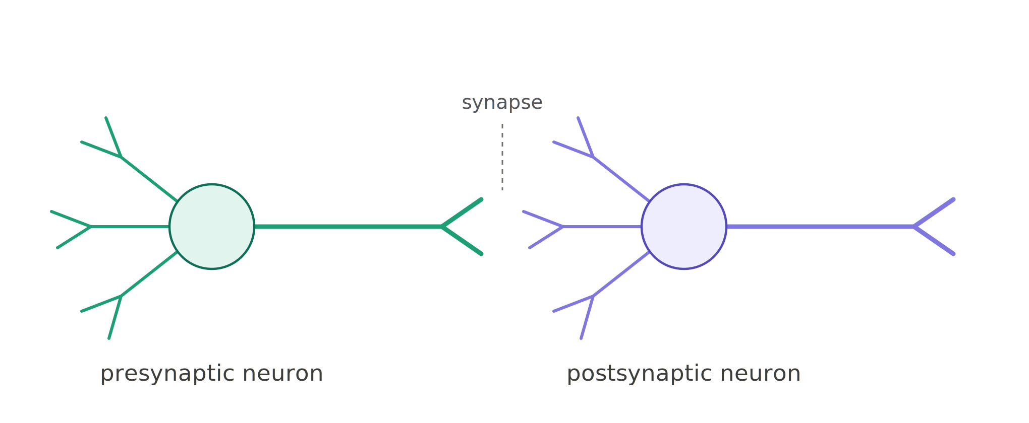
Now, in the diagram below, note which are the dendrites and which is the axon terminal.
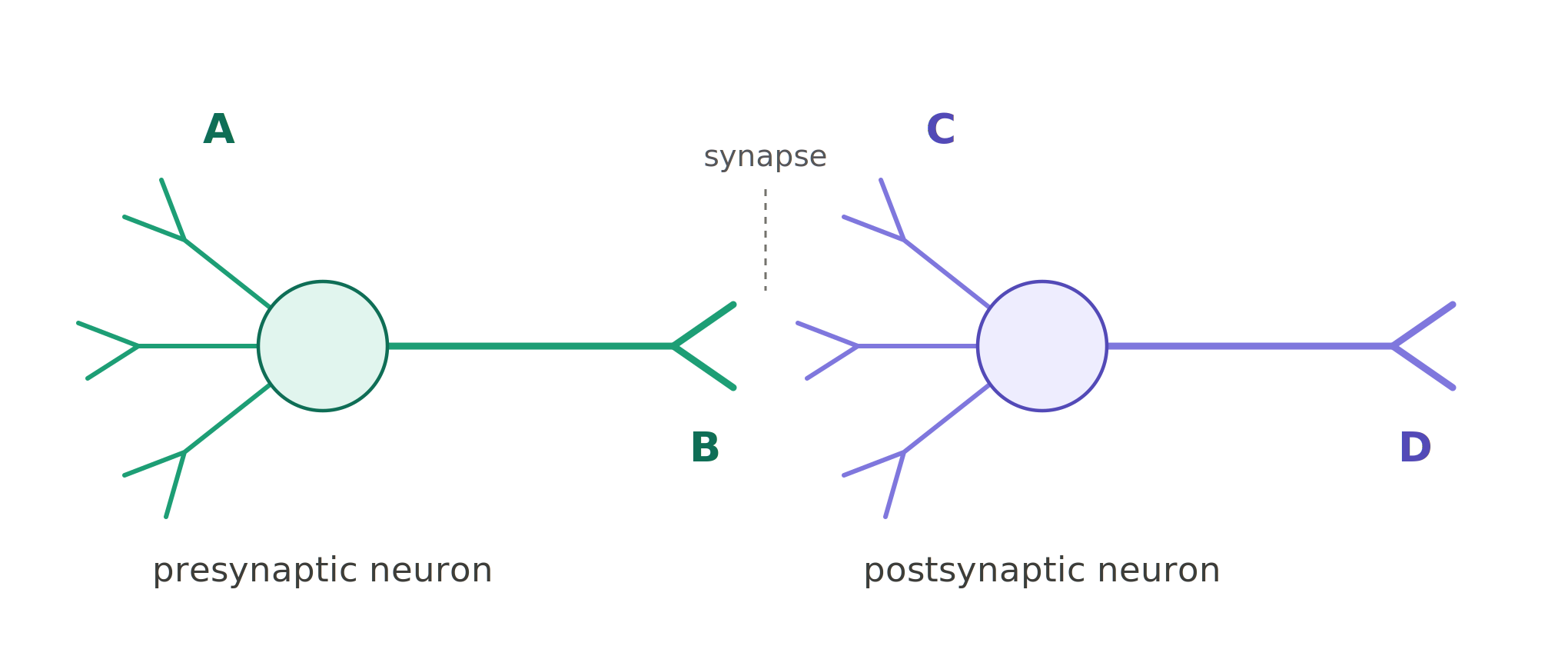
1.The axon terminal of the presynaptic neuron is labeled
2.The dendrites of the postsynaptic neuron are labeled
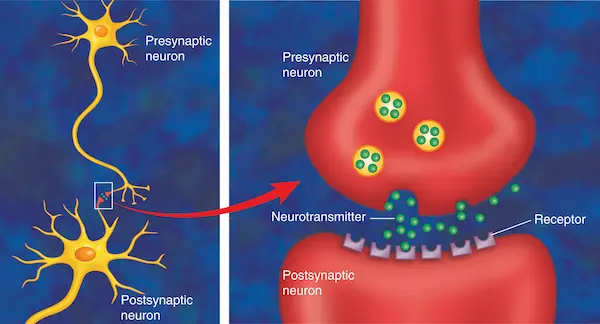
Notice in the illustration that neurotransmitter is released from an axon terminal of a presynaptic neuron, and binds to receptors located on a dendrite of a postsynaptic neuron. (Sometimes the receptors are on the cell body of the postsynaptic neuron.) Imagine that the presynaptic neuron has its dendrites in the skin, and that it was activated by the heat of a cup held in the hand. You can imagine Na+ channels opening in the dendrites and cell body, and if enough Na+ enters and the neuron is sufficiently depolarized, voltage-gated Na+ channels in the axon open, one after another, until neurotransmitter is released from the presynaptic terminal. The transmitter crosses the synapse and binds to receptors on the dendrites of the postsynaptic neuron. This causes opening of Na+ channels and inflow of Na+ into the dendrites. Again, if enough Na+ enters and the cell is sufficiently depolarized, the voltage-gated Na+ channels in the axon open, leading to release of neurotransmitter from that neuron.
Here are some missing pieces.
What does it mean for a neuron to fire, sending an action potential along its axon (section 2.2.4 and 2.2.5)? And when the action potential reaches the axon terminal, how is a neurotransmitter released (2.3.1, 2.3.2 and 2.3.3)? Finally, once the neurotransmitter is released from a presynaptic neuron, how does it excite (2.4.1) or inhibit (2.4.2) a postsynaptic neuron?
As you look into these questions, here are the key concepts you'll want to read about, in order of priority:
- Priority 1: EPSP, IPSP, hyperpolarization, neurotransmitter exocytosis, neurotransmitter receptor, neurotransmitter reuptake, neurotransmitter, degradation
- Priority 2: myelination, conduction velocity, spatial summation, temporal summation, agonist drug, antagonist drug
- Priority 3: ionotropic receptors, metabotropic receptors, second messengers
You can review these additional concepts using the Test Yourself Questions within chapter 2, and check your answers in the accompanying Student Resource page. If you don't have Principles..., you can still access the Test Yourself questions and answers at that link. From the same Student Resources page, anyone can access "Label the Figure" and Flashcard practice material relevant to these topics.
Happy studying.